What is it?
Calcific Tendonitis is a condition in which a small calcium deposit develops in the rotator cuff tendons causing the tissues in the subacromial space to become inflamed causing pain.
It is often difficult to distinguish between calcific tendonitis and subacromial pain however the treatment is the same.
The cause of calcific tendonitis is unknown but it tends to affect those in middle age.
What are the signs and symptoms?
Pain is felt in the region of the joint. This is usually a dull pain at night, which is made worse by lying on that side. Sharp pain is felt when reaching overhead,
There are two different presentations
1. Acute severe pain. This is usually described by patients as “the worst pain ever”. It comes on over a few hours and the patients will often attend A&E for pain relief. After the acute episode settles the milder form may become a feature.
2. Chronic, repeated episodes of moderate pain which is very similar to subacromial pain syndrome.
This can include
- Mild to severe pain
- Limited movement.
- Inability to hold the arm in certain positions e.g. reading a newspaper
The pain often has two forms
A continuous dull ache, which is often worse at night, is usually located over the lateral aspect of the shoulder joint and may extend someway down the side of the arm even as far as the elbow. Often it does not appear to be coming from the joint at all.
Sharp pain on lifting the arm is felt just below the acromion (the bony prominence at the top of the shoulder). The pain usually starts when the arm reaches horizontal and eases off if the arm can be brought all the way to vertical. This is the so called “painful arc”.
How is the diagnosis made?
The clinician will listen to the description of the symptoms and examine the shoulder. X-rays will be taken to examine the shape of the acromion and to help exclude other pathology within the shoulder. Ultrasound may also be used to identify the calcific deposit.

What is the initial treatment?
In the acute, severe phase a local anaesthetic and steroid injection into the subacromial bursa will produce rapid and lasting relief.
In the more chronic phase then the following the following may be tried.
- Avoidance of the aggravating activities.
- Anti-inflammatory drugs should be used and may be all that is required.
- Local injection of local anaesthetic and steroid often settle the acute episode.
- Ultrasound guided “barbotage” in which the calcium is needled and attempts are made to aspirate the deposit.
- Shockwave Therapy may also be recommended (as is used for Kidney Stones) to break up the deposit
What if the initial treatment fails?
If the symptoms do not improve then it may be necessary to surgically remove the joint. This is typically done using the arthroscope.
Arthroscopic excision of calcific deposits
Anaesthetic
General Anaesthetic with an interscalene block (Fully asleep with a local anaesthetic injection into the side of the neck will numb the nerves to the shoulder for post-operative pain relief)
Operation type
Arthroscopic – 3 0.5cm incisions, one at the back, one at the side and one at the front of the shoulder
Procedure
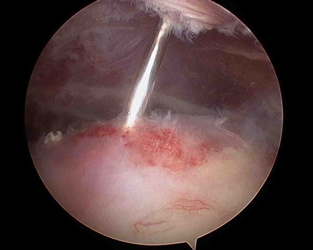
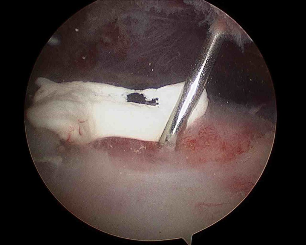
The gleno-humeral (shoulder) joint will be inspected first followed by the subacromial bursa and the rotator cuff. A soft tissue shaving device will be used to clear any scar tissue away and to remove any calcium

Needle in calcific deposit 
Calcific deposit
Wound Closure
Small butterfly paper stitches will be used to close the wounds.
Tegaderm waterproof dressings will be placed over the top of the paper stitches.
Pain relief
The anaesthetist will discuss a nerve block which will be administered after you are asleep, this means that you will wake up with no pain in the shoulder but the arm will feel numb for up to 12 hours. You will be prescribed painkillers to start taking when you get home and we encourage you to take these regularly for at least the first few days. There will be some discomfort but this settles quite rapidly and ice packs can be used in addition if you wish.
Wound care
The dressings will be changed before you go home and these can be left alone until they are removed. Typically they can be removed 10 days after surgery just by peeling them off and you do not need to visit the doctor for this.
The dressings are showerproof and you will be given some spares in case they start to peel off.
Rehabilitation after surgery
You will wake up with a sling on your arm. This can be removed as soon as you are comfortable, typically the next day.
You should try to use the arm as normally as possible, there will be some pain, particularly above shoulder height but you should not worry about this.
Surgical Risks
Every operation has a degree of risk. It is important that you are aware of these risks before you agree to proceed with your operation.
If you decide not to proceed with surgery there is a possibility that the symptoms will settle on their own but they may continue and they may get worse. You will not damage the shoulder if you decide not to proceed with surgery.
The most common or significant risks are outlined below. A risk of 0.1% means that 1 in 1000 people will suffer the complication
- Failure of the procedure to relieve symptoms: 5%
- Superficial Infection (requiring antibiotics): 0.16%
- Deep Infection (requiring further surgery): 0.02%
- PE (Pulmonary Embolus) (Blood clot in the lung): 0.13% – Blood thinning medication is required for several months. Can rarely result in death.
- DVT (Deep Vein Thrombosis) (Blood clot in the leg): 0.14% – Blood thinning medication is required for several months. Can lead to PE
- Nerve Injury: 0.01% – Usually temporary. Can cause weakness around the shoulder with loss of function and rarely can be permanent.
- Heart attack: 0.02%
- To put these numbers in perspective
The chance of:- Getting three balls in the UK national lottery: 0.9%
- Needing emergency treatment in the next year after being injured by a can, bottle, or jar: 0.1%
- Death by an accident at home: 0.01%
- For a 50 year old man in good health the 5 year risk of dying is 0.8%
Frequently asked questions
Return to work after surgery
This is very much dependent on the type of work that you do, whether you need to drive to get to work and the type of surgery that you have had done.
You, as the patient, have the best idea of the specific demands that are required of you to do your work safely and effectively.
Having an operation with an anaesthetic often takes more out of people than they would expect. Generally it is probably worth taking at least a week off from your regular work after having had any procedure.
You should discuss expected post-operative recovery and work with the surgeon before your operation.
Driving after surgery
To be able to drive safely you should be capable of actively moving your shoulder without assistance and without damaging the surgical repair. You should be able to react normally to avoid causing injury to yourself or others due to a lack of control.
Typically this is a no more than 2 weeks after surgery.
It is a UK requirement that, unless specific dispensation has been granted by the DVLA, a driver uses both arms to control the steering wheel.
It is the responsibility of the driver to ensure that they are in control of the vehicle at all times. They should be able to demonstrate this if stopped by the police.
It is not a requirement to notify the DVLA unless the medical conditions likely to affect safe driving persist for longer than three months after the date of the surgery.
Drivers must not drive under the influence of narcotic medications or within a minimum of 24 hours after an anaesthetic.
Although it is not essential, it may be wise to discuss your return to driving with your car insurance company.
Sports after surgery
You can start simple cardio such as walking or using a static bicycle immediately following surgery. Exercises which involve the shoulder can start as soon as you feel comfortable. Typically this is a few weeks but it may take 5 months or more for high demand overhead sports (swimming, tennis etc to return to normal)